Echocardiograms can detect aortic aneurysms, arrhythmias, cardiomyopathy, valve disease and heart failure. Also called a heart echo or just echo for short, an echocardiogram is an ultrasound of the heart.
Valley offers the full range of heart echocardiograms, including transesophageal, transthoracic and stress tests. We use 3D ultrasound images and ultrasound-enhancing agents to get the clearest picture of your heart.
Valley’s cardiac imaging program offers the highest level of accuracy and interpretation for echocardiograms. Our team also combines echoes with other diagnostic tests to get to the root of your heart condition.
With this information, your care team will tailor a care plan just for you.
What is an Echocardiogram?
A heart echocardiogram is a painless, noninvasive imaging test. An echocardiogram uses sound waves to create images of your heart and its structures. We may use it to assess:
- Aorta problems
- Blood clots
- Blood flow patterns
- Fluid buildup
- Heart size
- Heart pumping and relaxing function
- Heart valve function
- Pressure in the heart
Depending on the type of echo you need, we may use an ultrasound-enhancing agent during your echocardiogram. This improves the picture and gives us more accurate results.
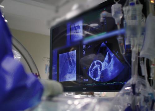
All types of echocardiograms use an ultrasound transducer and a computer. The transducer sends sound waves through the body, and the computer translates the sound waves into images.
Types of Heart Echocardiogram
There are different types of heart echocardiograms. The type of echo you’ll need depends on your symptoms or the heart condition we’re evaluating.
Transthoracic Echocardiogram (TTE)
A transthoracic echocardiography (TTE) is the most common type of heart echo. It’s used to assess your overall heart health. It can help identify the root of certain heart-related symptoms like shortness of breath and chest pain.
Here’s what you can expect during a TTE:
- You will lie on a table.
- If needed, your technician will give you an ultrasound-enhancing agent.
- The technician will apply gel to your chest and move the transducer over your skin to capture images of the heart.
- They may ask you to hold your breath or flip to your side.
- A TTE usually takes about 30 to 45 minutes to complete.
Transesophageal Echocardiography (TEE)
A transesophageal echocardiography (TEE) gathers more detailed images than the TTE by getting images through your esophagus. You may need a TEE if you have specific heart conditions, including heart valve conditions or blood clots.
Here’s what you can expect during a TEE:
- A provider will give you a sedative that allows you to sleep through the test.
- They will also numb your throat for your comfort.
- Once you are asleep, your provider will thread a small probe with a transducer down your esophagus.
- They will take images of your heart.
- The test usually takes about 45 minutes.
- After the test, we will monitor you for a few hours as you wake up from sedation.
Stress Echocardiogram
A stress echo, or stress test, uses transthoracic echocardiogram before and after exercise to assess your heart function. The stress test helps determine how well your heart is working.
We also perform dedicated studies to comprehensively evaluate hypertrophic cardiomyopathy (increased thickness of the heart muscle). And we use this test to evaluate valvular heart disease, such aortic stenosis and mitral valve regurgitation.
Here’s what you can expect during your stress test:
- Your technician will place electrodes that are attached to monitors on your chest.
- During the test, we’ll take images of your heart at rest.
- Then, you’ll walk on a treadmill for five to 10 minutes until your heart rate increases. We will then take images of the heart again.
- If you can’t use the treadmill, we can give you medicine that increases your heart rate like exercise.
- A stress echo can take about 30-60 minutes.
Echocardiogram Technologies at Valley
Valley uses advanced echo technologies that help us provide the most accurate cardiac imaging and diagnoses. These technologies include:
- 3D ultrasound allows us to see your heart structure heart function at different angles.
- Color doppler ultrasound shows your blood flow using different colors.
- Ultrasound-enhancing agent involves a solution given intravenously (IV) to highlight heart structures more clearly.
- Myocardial strain imaging detects early changes in heart muscle motion.
Why Choose Valley for Your Echocardiogram?
- Experience you can trust: Our cardiac imaging director is a cardiologist with subspecialty training in echocardiology and nuclear cardiology. This expertise means your cardiac imaging team is directly involved in your care planning. Your care team will use the echocardiogram results to best guide and personalize your treatment plan.
- Collaborative, team-based approach: Our cardiac imaging team works with Valley’s heart care specialists to determine the best treatment plan specific to your imaging results. We understand common and complex heart diseases and how they’re treated.
- Research focus: Cardiac imaging specialists at Valley actively lead research to help improve echocardiograms and our overall diagnostic capabilities. We also participate in clinical trials that incorporate advanced cardiac imaging to improve treatment for a wide range of heart diseases.
- Commitment to quality: Our cardiac program prides itself on having robust quality control measures. We continually review our imaging studies for consistency of imaging and reporting. This level of quality control provides you with the highest-quality images and allows your care team to determine the best treatment plan for your specific needs.
- Convenient imaging locations: Valley makes it as easy as possible to get an echocardiogram at several locations.


